


Dr Norman Nieder-Heitmann
MBChB (Stell), Dipl Ophth (SA), FC Ophth (SA), MMED Ophth (UCT)
Paarl Eye Centre
Tel: 021 871 1368
Age-related macular degeneration (ARMD) is one of the leading causes of blindness, accounting for almost 10% of cases worldwide. In developed countries, it is the most common cause of moderate to severe vision loss, and it is estimated that by 2040 more than 250 million people will suffer from this condition. Although it does not cause total blindness, it can severely affect central vision.
Due to advances in modern medicine, we now have a much better chance of living to an older age than our ancestors. Unfortunately, as the name indicates, advanced age is one of the main risk factors.
What is the retina and where is the macula?
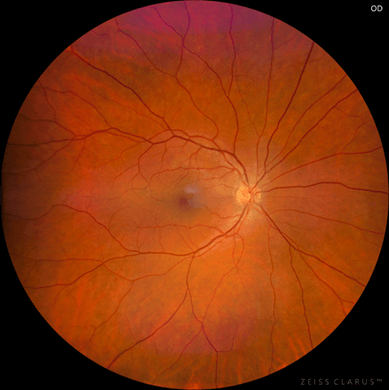
Image 1: A picture of the normal retina. The macula is circled in yellow with the central red circle marking the fovea.
The retina is a thin layer of brain tissue lining the inside of the eye. It consists of light-sensitive cells, known as rods and cones, which—together with numerous other nerve cells—organize our visual data. This information is then relayed via the optic nerve to the brain, where images are formed. In simple terms, the retina captures images, which are then processed and formed in the brain.
The macula is the central part of the retina responsible for sharp, central vision. This includes activities such as reading, writing, and watching TV. At the center of the macula lies the fovea, which allows us to perform the most detailed visual tasks like threading a needle.
What causes macular degeneration?
Macular degeneration is caused by a complex interplay of multiple risk factors that lead to irreversible damage of the nerve cells in the macula. The main risk factors are age (>50), smoking, and genetic predisposition. Other risk factors include Caucasian race, female sex, blue eyes, obesity, high blood pressure, and elevated cholesterol levels.
Macular degeneration can be divided into two main types: dry ARMD and wet (neovascular) ARMD. The dry type is more common and progresses slowly and gradually, whereas the wet type is less common but more serious, with rapid and aggressive progression.
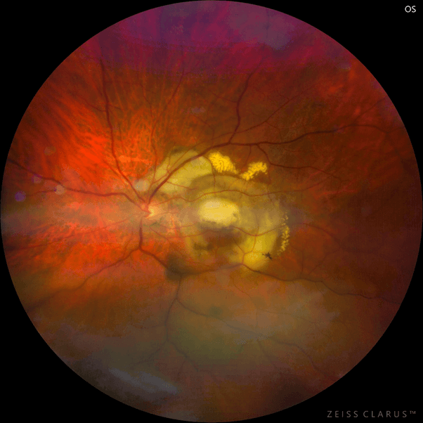
Image 2: Dry macular degeneration
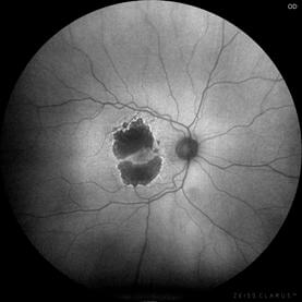
Image 3: Fundus autofluorescence demonstrates the area affected in black

Image 4: A severe form of wet macular degeneration which presents a large bleed in the macula
Image 5: An example of the final scarring of the macula in an untreated case with wet macular degeneration
Symptoms of macular degeneration.
Common symptoms of ARMD include blurry or fuzzy vision, dark spots in the center of vision, distortion of straight lines, and difficulty recognizing faces or reading fine print.
Treatment and management.
It is extremely important to manage modifiable risk factors such as smoking, excess weight, high blood pressure, and high cholesterol levels. Large studies (AREDS2 clinical trials) have shown that antioxidant and mineral supplementation can reduce the risk of progression in people with established ARMD. Ocuvite is a fixed-combination supplement that is taken twice daily. A diet rich in leafy green vegetables and omega-3 fatty acids also provides many of these beneficial vitamins and nutrients.
For advanced dry macular degeneration with established geographic atrophy, a newly approved injection called Pegcetacoplan has been shown to reduce the size of the affected area. Although not yet approved in South Africa, it offers hope to patients with dry ARMD.
Wet macular degenerationis treated with medications called anti-VEGFs, which are injected directly into the eye (e.g., Aflibercept, Ranibizumab, Faricimab). Treatment should be started as early as possible to maximize the chance of preserving vision. This is an exciting and fast-evolving field in ophthalmology, with extensive ongoing research.
For patients with irreversible central vision loss, low-vision aids are available to help them carry out daily activities and improve their quality of life. These include tools such as magnifiers, electronic readers, and adaptive lighting.
